
StraightSmile Solutions®
The EBITDA Playbook for Adding Interceptive Ortho to a Pediatric Dental Practice
The EBITDA Playbook for Adding Interceptive Ortho to a Pediatric Dental Practice
Introduction
Dr. Amanda of Straight Smile Solutions explains how adding interceptive orthodontics can significantly increase EBITDA in pediatric and primary care dental practices. By integrating early orthodontic services, especially with clear aligners and simple interceptive approaches, practices can grow profitability, reduce overhead, and improve long-term practice valuation. When implemented strategically, orthodontics becomes a high-margin service that strengthens both daily revenue and future exit opportunities.
Why Orthodontics Improves EBITDA
- Orthodontics adds revenue without requiring significant capital investment
- Minimal need for loans, borrowing, or depreciating equipment
- High profit potential with limited marketing and operational costs
- Improves overall practice value for future sale opportunities
Low-Overhead, High-Profit Implementation
- Best systems are simple and efficient
- Requires minimal staff training and doctor chair time
- Clear aligners and interceptive orthodontics fit easily into existing workflows
- Designed to maximize income without increasing operational complexity
The Market Gap in Early Interceptive Care
- Most orthodontists focus on teens and adults
- Limited focus on early interceptive treatment for children under nine
- Early orthodontic needs are often underserved in many communities
- Pediatric dental practices are uniquely positioned to fill this gap
Why Phase One Interceptive Orthodontics Makes Sense
- Treating problems early prevents complex orthodontic cases later
- Growth modification, expanders, airway management, and habit correction are more effective in young patients
- Early care reduces the severity of future orthodontic challenges
- Builds long-term patient trust and retention within the practice
Team Readiness and Practice Culture
- Pediatric teams already have the patience and environment needed for young patients
- A fun, calm practice culture makes early orthodontics easier to deliver
- Confidence and training are key barriers, not clinical difficulty
Preparing for Practice Sale
- Orthodontics strengthens EBITDA when cases are completed properly
- Starting many unfinished cases can negatively affect practice valuation
- Strategic case completion improves buyer confidence
Conclusion
Dr. Amanda emphasizes that interceptive orthodontics is one of the most effective and cost-efficient ways to increase profitability in pediatric dental practices. With low overhead, strong demand, and minimal competition, early orthodontic care enhances EBITDA while improving patient outcomes. When done correctly, it positions a practice for sustainable growth and a stronger future sale value.
CSA vs OSA (Sleep Apnea): Which Needs MMA—and Which Can Be Treated With MARPE, MSE, and Orthodontics?
Introduction
Dr. Amanda from Straight Smile Solutions breaks down a critical concept many dentists misunderstand: not all sleep apnea is the same, and not all cases can be treated orthodontically. With obstructive sleep apnea (OSA) treatment becoming increasingly common in dentistry, it’s essential to understand when orthodontic expansion can help and when only medical or surgical intervention is appropriate.
- Dentistry’s Role in Sleep Apnea
- Dentists are expected to screen, understand, and refer sleep apnea cases appropriately.
- Treating the root cause early, especially through proper interceptive orthodontics in growing children, can significantly reduce future airway problems.
- OSA vs CSA: Know the Difference
- Obstructive Sleep Apnea (OSA):
- Caused by a physical blockage of the airway.
- Common contributors: small jaws, tongue position, obesity, large neck circumference, tonsils/adenoids.
- Patients struggle to breathe against obstruction; often loud and common.
- This is the only type of dentistry that can potentially help.
- Central Sleep Apnea (CSA):
- A neurological issue, not an obstruction.
- Associated with heart failure, stroke, opioid use, or high altitude.
- Rare and not treatable by dentistry or orthodontics.
- Requires medical or airway-focused surgical management.
- Obstructive Sleep Apnea (OSA):
- Limits of MARPE, MSE, and Similar Appliances
- These appliances mainly address transverse (width) deficiencies only.
- They do not create forward jaw growth or correct vertical or AP discrepancies.
- If a recessed mandible causes apnea, expansion alone will not help.
- Misuse can lead to tipped teeth, bite damage, and unstable results.
- When MMA (Jaw Surgery) Is Necessary
- Required when airway issues are 3D (vertical + AP + transverse).
- MMA moves both jaws forward, enlarging the airway and repositioning soft tissues.
- Patients with deep bites, skeletal asymmetries, or significant jaw discrepancies typically need surgery followed by orthodontics for proper function and esthetics.
Conclusion
Understanding whether a patient has OSA or CSA is non-negotiable. Orthodontic expansion can help only in very specific obstructive cases with isolated maxillary constriction. When the airway problem is skeletal or multidimensional, MMA surgery, not appliances, is the predictable solution. Proper diagnosis, collaboration with MDs, and respecting orthodontic limits are key to ethical and effective sleep apnea care.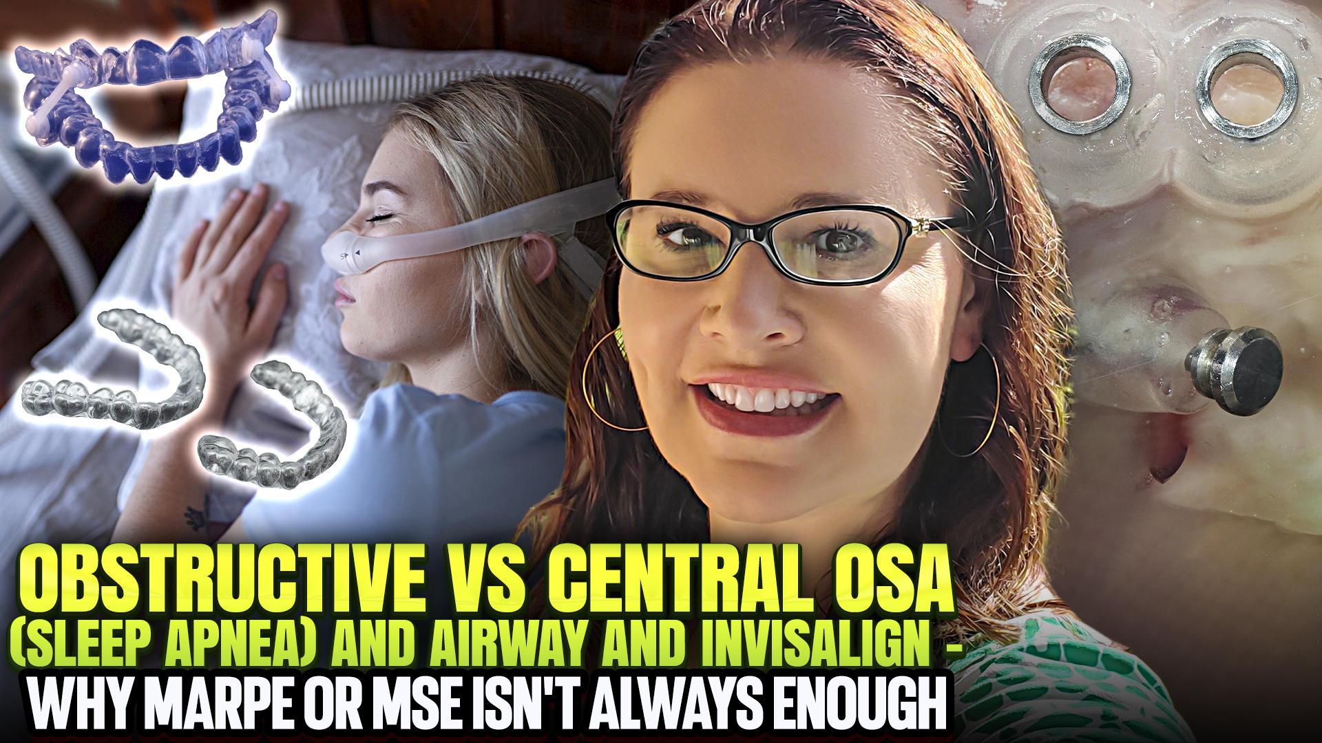 CSA vs OSA (Sleep Apnea): Which Needs MMA—and Which Can Be Treated With MARPE, MSE, and Orthodontics?
Thin Bone vs Invisalign A.I. and Fenestrations: Using Analog Techniques to Get Better ClinChecks
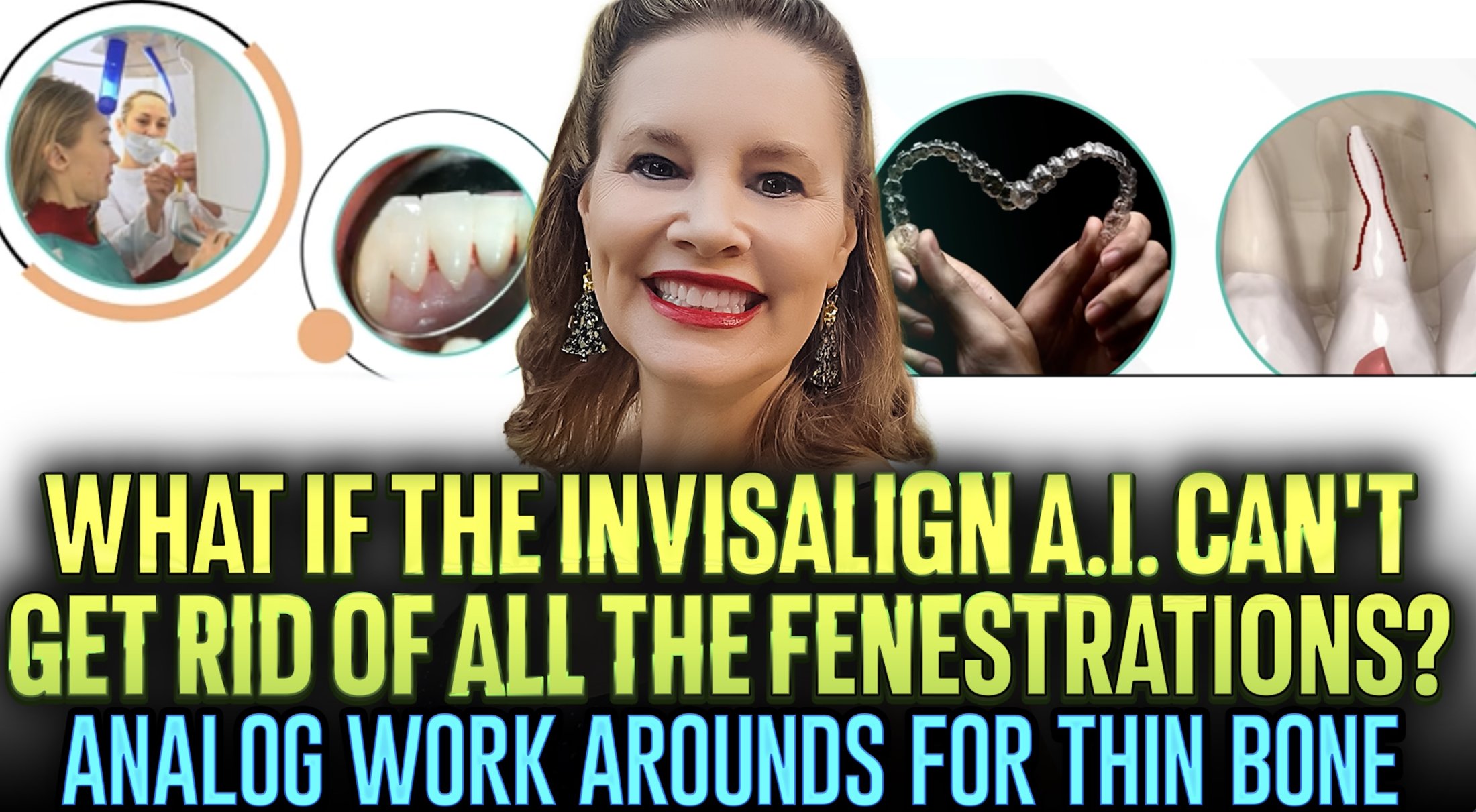 Thin Bone vs Invisalign A.I. and Fenestrations: Using Analog Techniques to Get Better ClinChecks
Introduction
Dr. Amanda of Straight Smile Solutions discusses how Invisalign’s AI-driven ClinCheck and CBCT integration can sometimes flag root fenestrations or thin bone concerns that may not fully reflect real-world biological outcomes. While AI tools are powerful, she emphasizes that they are decision-support systems, not substitutes for clinical judgment. Understanding how to interpret these warnings and apply analog orthodontic principles is critical for predictable, safe treatment planning.
Understanding Invisalign AI and CBCT Integration
AI segments roots and alveolar bone from CBCT scans
Creates a 3D model that moves roots and crowns together
Highlights biological limits based on existing bone anatomy
Red warnings often represent worst-case scenarios, not certainties
Why Fenestrations Appear in ClinChecks
AI works within the current bony housing and does not predict future bone adaptation
Thin bone may be difficult for AI to visualize accurately
Over-torquing roots without adequate crown movement can trigger warnings
Algorithms may overcorrect in pursuit of root parallelism
The Role of Slow, Analog Tooth Movement
Very slow, deliberate movement increases osteoplastic activity
Constant, low forces favor bone build over breakdown
Slowing movements two to four times beyond default staging may reduce risk
Arch widening and controlled proclination may be tolerated biologically
Clinical Adjustments Before Abandoning a Case
Adjust torque before increasing IPR
Keep movements within existing bony housing when possible
Be precise and deliberate in instructions to the technician
Accept that some fenestrations cannot be fully engineered away
Risk Management and Interdisciplinary Planning
Loop periodontists into high-risk cases early
Obtain periodontal approval in writing
Monitor closely throughout treatment
Be prepared to discontinue treatment if risks outweigh benefits
Conclusion
Dr. Amanda emphasizes that Invisalign AI is a powerful visualization tool, but not a replacement for sound orthodontic judgment. Thin bone and fenestration warnings should prompt thoughtful planning, slower movements, and interdisciplinary collaboration. By combining modern AI insights with analog orthodontic principles, clinicians can produce safer, more realistic ClinChecks while protecting both patients and practices.
Lip Incompetence, Mentalis Strain, and Class II High-Angle Ortho Patients – The Airway Trifecta

Introduction
Dr. Amanda of Straight Smile Solutions explains the close relationship between lip incompetence and mentalis strain, a combination frequently seen in Class II, high-angle orthodontic patients. Lip incompetence refers to the inability to maintain a closed mouth posture at rest, often forcing the chin muscle (mentalis) to work overtime. Understanding which condition comes first and how they interact is critical for proper orthodontic diagnosis, treatment planning, and long-term stability.
- Definition of lip incompetence
- Inability to keep lips gently closed at rest without effort
- Often accompanied by visible mentalis strain
- Role of the mentalis muscle
- Overactive mentalis attempts to force the lips together
- Leads to chin dimpling or a “peach-pit” texture, especially noticeable in women
- Underlying growth issues
- Occurs when jaws, soft tissue, or muscles are not proportionally developed
- Asymmetry or underdeveloped structures increase the effort needed to close the mouth
- Adult vs. pediatric considerations
- Limited correction options in adults
- Myofunctional therapy and orthodontics may help, but jaw surgery is often required
- Early intervention in children can significantly improve outcomes
- Impact on orthodontic treatment
- Excessive chin and lip pressure can disrupt tooth movement
- External forces act similarly to thumb sucking or ill-fitting appliances
- Can prevent proper orthodontic alignment if not addressed first
- Dental and facial consequences
- Increased risk of tooth over-eruption
- Vertical growth patterns and long-face appearance
- Narrow palates, recessed chins, and gummy smiles
- Risk of relapse
- Unresolved mentalis strain greatly increases post-treatment instability
Conclusion
Lip incompetence and mentalis strain are not cosmetic concerns as they directly affect orthodontic mechanics, facial growth, airway considerations, and treatment stability. Dr. Amanda emphasizes the importance of identifying and addressing these issues early, particularly in growing patients. For orthodontists, recognizing mentalis strain and factoring it into the treatment plan is essential to achieving predictable, stable, and healthy results.
The 60-Day Blueprint for Launching Orthodontics in a Pediatric Dental Practice
I. Introduction
Dr. Amanda from Straight Smile Solutions outlines a practical 60-day roadmap for integrating orthodontics into a pediatric dental practice. The approach works for both de novo and existing practices, with de novo offices having a natural advantage. Early orthodontic and airway screening is not optional; it aligns with AAPD and ADA standards of care. Offering orthodontics in-house improves continuity of care, conversion rates, and long-term patient outcomes.
II. Why Pediatric Practices Are Ideal for Orthodontics
- Pediatric dentists already see patients at the ideal growth window for interceptive orthodontics.
- Parents expect guidance on growth, airway, and development, not just cavities.
- When orthodontic screenings are standard from day one, parents view them as routine, not sales-driven.
- Referral-only models often fail due to poor follow-through and lack of interdisciplinary communication.
III. Screening, Referrals, and Conversion Rates
- Every child must be screened regularly for orthodontic and airway issues.
- If a child needs treatment and it’s explained correctly, conversion rates should exceed 80–90%.
- Simply referring and “checking a box” is not enough; follow-up must occur at every recall visit.
- Orthodontists should provide clear reports explaining what is being done and why.
IV. Learning Orthodontics the Right Way
- Orthodontics is not mastered through short courses or rigid templates.
- True competency comes from repetition, pattern recognition, and mentorship, similar to residency training.
- Growth, airway, genetics, and facial development must all be understood together.
- AI and modern tools make orthodontics more efficient, but clinical judgment remains critical.
V. Essential Tools to Get Started
- An iTero scanner is essential for pediatric orthodontics, new or refurbished.
- Clear aligners and digital workflows simplify treatment and increase efficiency.
- Braces and expanders remain effective, but aligners improve flexibility during mixed dentition.
Conclusion
Launching orthodontics in a pediatric dental practice within 60 days is both realistic and ethical when done correctly. By standardizing screenings, investing in the right tools, and committing to proper education, pediatric dentists can dramatically improve patient care while building a sustainable, profitable orthodontic service that supports long-term facial growth and airway health.
How to Future-Proof Your Practice and Make an Extra $100K per Year with Orthodontics and A.I.
How to Future-Proof Your Practice and Make an Extra $100K per Year with Orthodontics and A.I.
I. Introduction
Dr. Amanda from Straight Smile Solutions discusses how orthodontics has evolved in 2026 and beyond. Many practices can add $100,000+ in annual revenue without major new investments. The key lies in optimizing existing systems, staff, workflows, and smart use of A.I. When done correctly, orthodontics becomes both financially rewarding and professionally fulfilling.
II. The Reality of Modern Orthodontics in 2026
- Most practices already have what they need to scale orthodontics.
- Success depends on choosing the right orthodontic services, not buying the most equipment.
- Doctors who follow proven systems often exceed revenue expectations while improving patient satisfaction.
- Orthodontics can become one of the most enjoyable and meaningful parts of clinical dentistry.
III. The Role of A.I. in Orthodontic Efficiency
- A.I. is a support tool, not a decision-maker; clinical judgment remains essential.
- The doctor retains full responsibility for diagnosis and outcomes.
- A.I. is best used to streamline workflows, reduce time, and improve consistency, not replace expertise.
IV. Where A.I. is Already Transforming Orthodontics
- Cephalometric analysis
- Eliminates manual tracing and saves significant time
- Helpful even for experienced clinicians as a confirmation tool
- Digital treatment planning
- Clear aligners, indirect bonding, and setup simulations are largely A.I.-driven
- Requires careful review and communication with technicians
- Malocclusion detection tools
- Useful for less experienced providers
- Optional for seasoned orthodontic clinicians
- Outcome simulations & appliance design
- Invisalign and other systems already rely heavily on A.I.
- Enhances patient understanding and case acceptance
- Virtual monitoring
- Reduces unnecessary in-office visits
- Saves chair time, increases efficiency, and improves patient satisfaction
V. Rethinking Traditional Braces and Old Workflows
- Monthly in-person visits are outdated and inefficient.
- Traditional braces with frequent adjustments increase:
- Chair time
- Gum inflammation
- Bone stress and root resorption risk
- Modern orthodontics prioritizes lighter forces and smarter monitoring.
VI. Equipment: What You Actually Need (and What You Don’t)
- An iTero scanner is highly recommended, but it does not need to be new.
- Older models work well for orthodontics and save tens of thousands of dollars.
- Supplies depend on your orthodontic menu:
- Removables: minimal additional costs
- Phase 1 ortho: very low overhead
- Fixed appliances: modest investment (bands, wires, basic instruments)
- Pediatric dentists may benefit from additional scanning or imaging technology, but it’s not mandatory.
VII. Why $100K Is Just the Starting Point
- Adding $100,000 annually is very achievable with proper systems.
- Practices that fully integrate orthodontics often exceed this benchmark.
- The biggest limiting factor is process, not equipment.
- With the right structure, orthodontics becomes scalable, ethical, and predictable.
Conclusion
Orthodontics combined with thoughtful A.I. integration offers a powerful way to future-proof your practice. By modernizing workflows and avoiding over-investment in equipment, doctors can increase revenue, reduce inefficiencies, and deliver better care. As Dr. Amanda emphasizes, when orthodontics is done the right way, everyone wins: the practice, the patients, and the provider.
The Dangers of Overusing Reverse Curve of Spee (RCS) Wires in Braces or Using Them at the Wrong Time
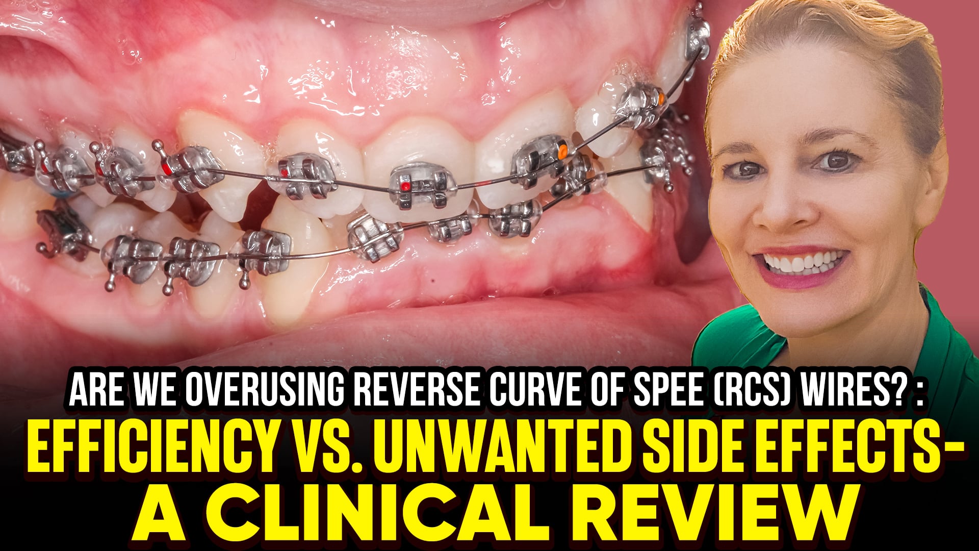 The Dangers of Overusing Reverse Curve of Spee (RCS) Wires in Braces or Using Them at the Wrong Time
Introduction
Dr. Amanda of Straight Smile Solutions addresses the common misuse of Reverse Curve of Spee (RCS) wires in fixed orthodontic treatment. This discussion focuses specifically on lower-arch RCS wires, not accentuated Curve of Spee wires. RCS wires are often misunderstood, overused, or applied prematurely, leading to unintended side effects. Dr. Amanda emphasizes that this is an advanced topic and should be considered only after mastering straight-wire fundamentals.
When RCS Wires Become a Problem
Why RCS Wires Are Risky
Considered a “cheat” or “lazy” wire in traditional orthodontic training.
They can create uncontrolled forces, leading to unexpected reactions elsewhere in the dentition.
Improper use often opens “a can of worms” rather than solving the underlying problem.
Common Mistakes
Using RCS wires too early, before:
Full alignment and leveling
Transverse and AP correction
Root parallelism confirmed on pano
Assuming all deep bites are the same:
Upper incisor over-eruption
Lower incisor over-eruption
Posterior up righting issues
Skeletal deep bites (not orthodontically correctable)
Better Alternatives (Often Preferred)
Fixed bite plates
Intrusion arches
Bias bracketing
Posterior box elastics with bite turbos
These approaches offer more controlled and predictable biomechanics.
When RCS May Be Appropriate
Only after:
Teeth are straight (2nd molar to 2nd molar)
Overjet and spacing are ideal
Roots are parallel on pano
Used for a mild residual deep bite caused by a lower Curve of Spee.
Must be worked up gradually
Critical Safety Check
Assess for lower anterior blanching before placement.
If blanching is present, RCS use risks pushing incisors out of the bone.
Once dehiscence occurs, it cannot be reversed and may involve periodontium.
Conclusion
Reverse Curve of Spee wires are not a shortcut; they are a precision tool with narrow indications. When used too early or without proper diagnosis, they can cause irreversible damage. Proper sequencing, diagnosis, and respect for biomechanics are essential to avoid turning a small bite issue into a major problem.
Pain and Discomfort Management in Dentistry: Teeth Pain, Braces, RPE, and Invisalign
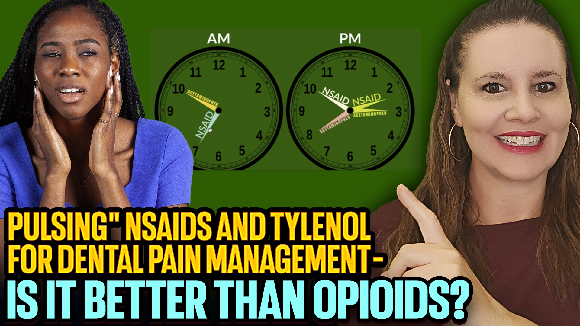
I. Introduction
- Dr. Amanda from Straight Smile Solutions outlines her standard protocol for managing discomfort during orthodontic treatment.
- Most patients do well with basic over-the-counter pain relief, but some are more sensitive and require a structured approach.
- This protocol is commonly used for braces, Invisalign/aligners, RPEs, and general dental discomfort.
- The goal is to control pain effectively while remaining safe and conservative.
II. Pain Management Protocol
- General Philosophy
- Pain management is discussed in detail only when patients express anxiety or discomfort.
- Treatment is short-term and typically limited to 2–3 days.
- Patients should always consult their physician before starting any medication protocol.
- Medication Categories
- NSAIDs (anti-inflammatory, kidney-metabolized):
- Examples: Motrin, Advil, Aleve, Aspirin
- Reduce inflammation associated with tooth movement
- Acetaminophen (pain reliever, liver-metabolized):
- Example: Tylenol
- Works through a different pain pathway than NSAIDs
- NSAIDs (anti-inflammatory, kidney-metabolized):
- Why Pulsing Works
- NSAIDs and acetaminophen can be used together but not at the same time.
- Alternating them (“pulsing”) improves pain control.
- Keeps pain blocked before medication wears off.
- Safety Guidelines
- Never exceed the maximum daily dose.
- Always take medications with food, especially NSAIDs.
- Dosage varies based on age, weight, and health status.
- Avoid NSAIDs with kidney issues; avoid acetaminophen with liver disease.
- Timing for Orthodontic Treatment
- Invisalign / Aligners:
- Start 1 hour before tray change or immediately after.
- Continue for 2–3 days while using chewies.
- Most patients do not need medication every tray.
- Braces or RPE Adjustments:
- Start 1 hour before activation.
- Continue for several days post-adjustment.
- Gradually taper off as discomfort improves.
- Invisalign / Aligners:
- Medication Duration
- NSAIDs last approximately 6–8 hours.
- Acetaminophen lasts 4–6 hours.
- Alternating doses maintains consistent pain relief.
III. Conclusion
Dr. Amanda’s pulsed pain management approach provides safe, effective, short-term relief for orthodontic discomfort. By alternating NSAIDs and acetaminophen appropriately and timing medication around orthodontic adjustments, patients can minimize pain while avoiding unnecessary medication use. As always, individualized care and physician guidance are essential for optimal outcomes.
Could Your Unilateral POB, Can’t, or Asymmetry Be Related to Menopause? Understanding Condylar Hypoplasia
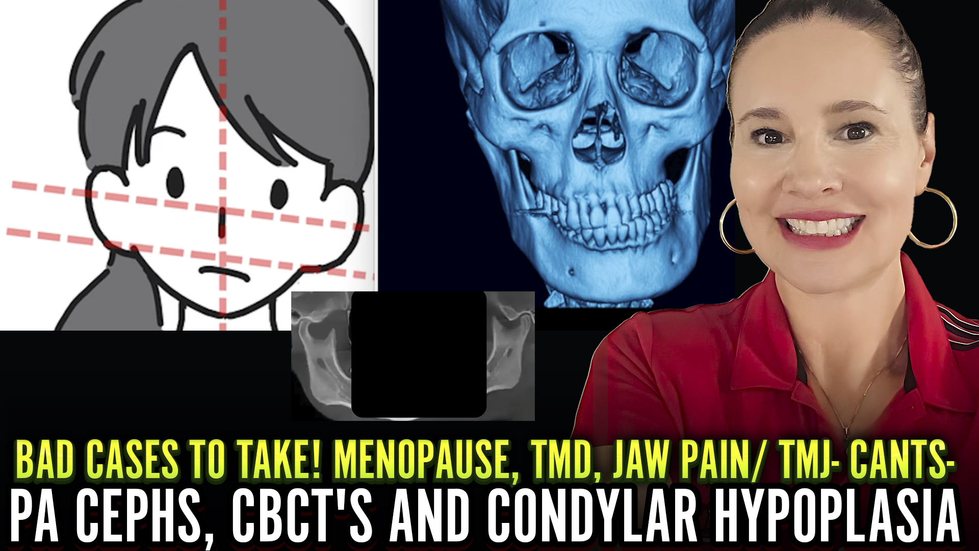
I. Introduction
- Dr. Amanda from Straight Smile Solutions discusses a commonly overlooked cause of adult-onset bite asymmetries.
- These issues are most often seen in adult patients in their 40s–60s, particularly females.
- New-onset unilateral posterior open bites (POB), cants, or facial asymmetries may not be orthodontic in origin.
- In some cases, the root cause lies in TMJ pathology, specifically condylar hypoplasia or condylar resorption, rather than teeth or jaw alignment alone.
II. When Orthodontics Is Not the Answer
- Clinicians should avoid immediately focusing on:
- Teeth
- Braces or aligners
- Jaw position
- Facial esthetics
- Instead, consider joint health and the possible need for joint replacement.
- Adult patients who previously had stable bites but now show progressive asymmetry warrant deeper investigation.
III. What Is Condylar Hypoplasia?
- Condylar hypoplasia is a condition where the mandibular condyle is smaller, underdeveloped, or resorbing.
- It can be:
- Congenital (rare, ~1 in 5,600 births)
- Acquired due to trauma, infection, inflammation, or systemic factors
- Progressive changes in condylar size or shape can cause:
- Facial asymmetry
- Bite canting
- Unilateral or bilateral posterior open bites
IV. The Role of CBCT and Modern Imaging
- In the past, 2D panos made it difficult to distinguish true pathology from artifacts.
- With CBCT and radiology reports, clinicians can now clearly assess:
- Condylar size and shape
- Side-to-side asymmetry
- Signs of resorption or degeneration
- Any adult patient with unexplained POBs or cants should have focused condylar analysis included in imaging reports.
V. Menopause, Hormones, and TMJ Changes
- Hormonal shifts during perimenopause and menopause can significantly affect bone and joint health.
- A sharp drop in estrogen, especially without hormone replacement therapy (HRT), may:
- Increase inflammation
- Accelerate bone resorption
- Worsen TMJ degeneration
- Exacerbate existing asymmetries
- Menopause can cause latent issues to suddenly become clinically obvious and progressive.
VI. Clinical Implications for Providers
- New-onset adult asymmetry should raise red flags before starting aligner therapy.
- Recommended steps include:
- Ordering CBCT with condylar evaluation
- Requesting radiologist commentary on TMJ pathology
- Avoiding orthodontic camouflage
- These cases often require jaw surgery and condylar replacement, followed by orthodontics, not the reverse.
Conclusion
Adult posterior open bites, cants, and asymmetries, especially in peri- or postmenopausal patients, may signal underlying condylar pathology rather than orthodontic relapse. Recognizing the influence of hormonal changes and joint degeneration is critical. Proper diagnosis protects patients from ineffective treatment and ensures they are guided toward the appropriate surgical and interdisciplinary care pathway.
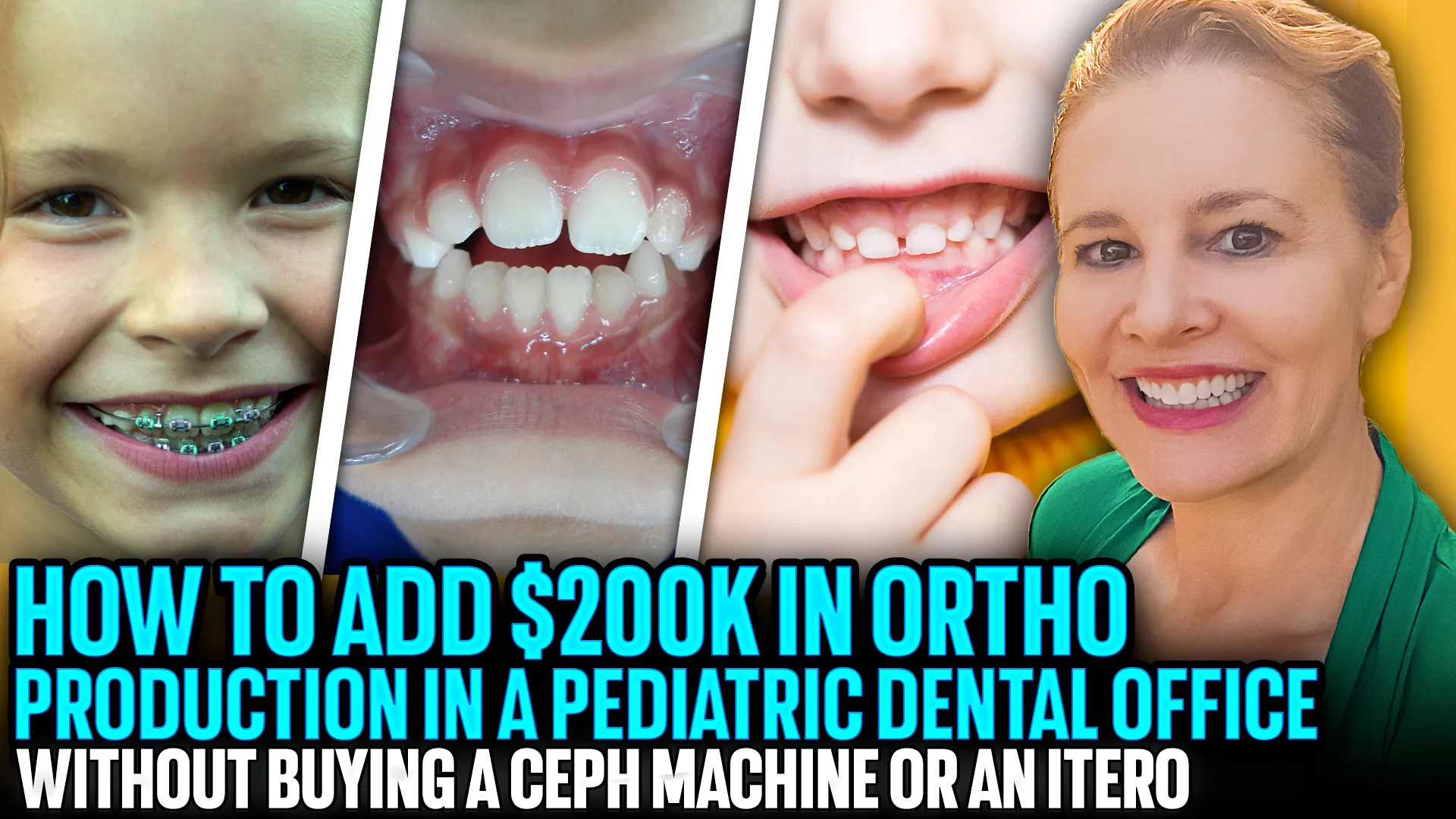
Adding $200K in Orthodontic Production to a Pediatric Dental Office Without a Ceph or iTero
I. Introduction
- Dr. Amanda from Straight Smile Solutions explains how pediatric dental offices can add $200,000 in orthodontic production in 2026 without purchasing a ceph machine or iTero scanner.
- This strategy is designed for pediatric dentists who already have a CBCT with a large field of view (FOV) and want to implement interceptive orthodontics efficiently.
- The key is early screening, systems, and team-driven workflows, not expensive new equipment.
II. Start with Early Screening: “Read the 2–5s”
- Every pediatric practice already has future orthodontic patients—you just need to identify them.
- By evaluating facial profile, lips, smile, and posture, orthodontic needs can often be predicted in children as young as 2–5 years old, even without X-rays.
- Early indicators include:
- Constricted palates or jaws
- Airway and mouth-breathing issues
- Myofunctional problems
- Vertical or transverse discrepancies
- Shifts, trauma, or palatal impingement
III. Build a Queue, Not Just a Case
- Place identified patients into a tracking or recall system and revisit the conversation at every visit.
- Discuss contributing factors such as:
- Nasal breathing and airway health
- Diet and posture
- ENT or myofunctional referrals
- Early habit correction tools
- While these visits may not always be billable orthodontically, they set up high-value Phase 1 and Phase 2 cases later.
IV. Why a CBCT Is Enough
- A CBCT with a sufficiently large FOV allows for:
- Pano and airway evaluation
- PAS assessment
- Growth and development monitoring
- A ceph is not mandatory, and while iTero improves efficiency, it is not required to start generating revenue.
- Interceptive orthodontics can be designed to be team-driven, especially with removable and simplified systems.
Conclusion
Adding $200K in orthodontic production does not require new machines it requires intentional screening, early conversations, and systems-based interceptive care. By identifying problems early and guiding families through proactive or reactive choices, pediatric dental offices can ethically grow orthodontic services while improving long-term patient outcomes.