Introduction
Dr. Amanda explains how to monitor patients transitioning from Phase 1 to Phase 2 orthodontic treatment, with a focus on preventing canine impaction. This guidance also applies to patients placed on observation when Phase 1 is not yet indicated. The goal is to understand monitoring frequency, eruption checkpoints, and how panoramic X-rays guide decision-making as canines navigate their eruptive paths.
- Purpose of Phase 1 Treatment
- Phase 1 exists to get patients out of trouble early using interceptive strategies.
• Primary goals: treat transverse discrepancies, vertical problems, and AP/sagittal issues.
• Examples include open bites, deep bites, anterior/posterior crossbites, overjets, negative overjets, and functional shifts.
• Phase 1 also aims to create the correct arch shape and adequate space for permanent teeth—improving eruptive paths and reducing impaction risks.
• Canines pose the highest impaction risk; creating proper arch form and space helps them self-correct without surgical or extraction intervention.
- Monitoring Canine Eruption with Panoramic X-Rays
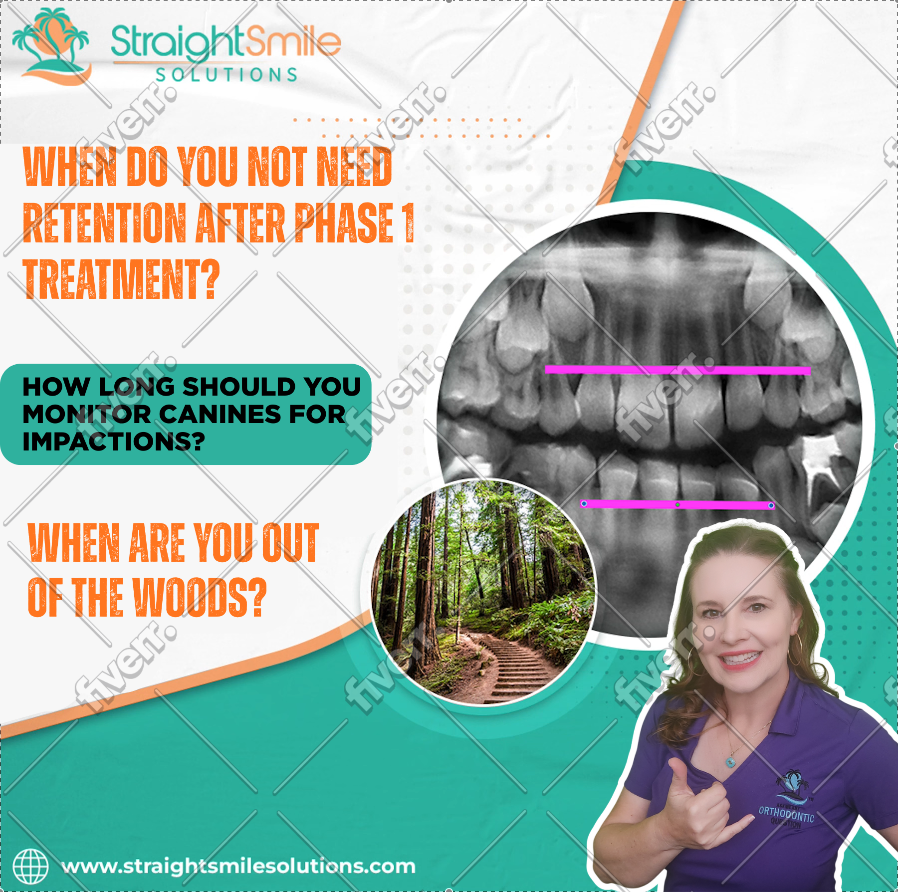
- If an initial panoramic at age 7–8 shows concern, monitor at least annually, and every 6 months if the problem is significant.
• Avoid unnecessary radiation: combine palpation of canine bulges with visual monitoring to reduce exposure.
• Key target: ensure canines are progressing past the height of contour of the maxillary incisors (approximately teeth #7–10).
• Before crossing this contour, canines remain at risk of getting hung up; after passing it, eruption is generally predictable and safe.
• Vertical orientation of the canine root and crown indicates a healthy eruptive path.
- Transitioning Safely into Phase 2
- Once canines clear the height-of-contour threshold, the risk of impaction drops sharply.
• A smooth transition into Phase 2 is expected when spacing and arch form are handled correctly in Phase 1.
• Phase 2 should be straightforward, with minimal alignment or bite correction needed.
Conclusion
Consistent monitoring during the Phase 1–Phase 2 period, especially of the maxillary canines, is essential for preventing impaction. Strategic timing of panoramic X-rays, careful palpation, and understanding of eruption landmarks ensure safe and predictable outcomes, as well as easier Phase 2 treatment.