
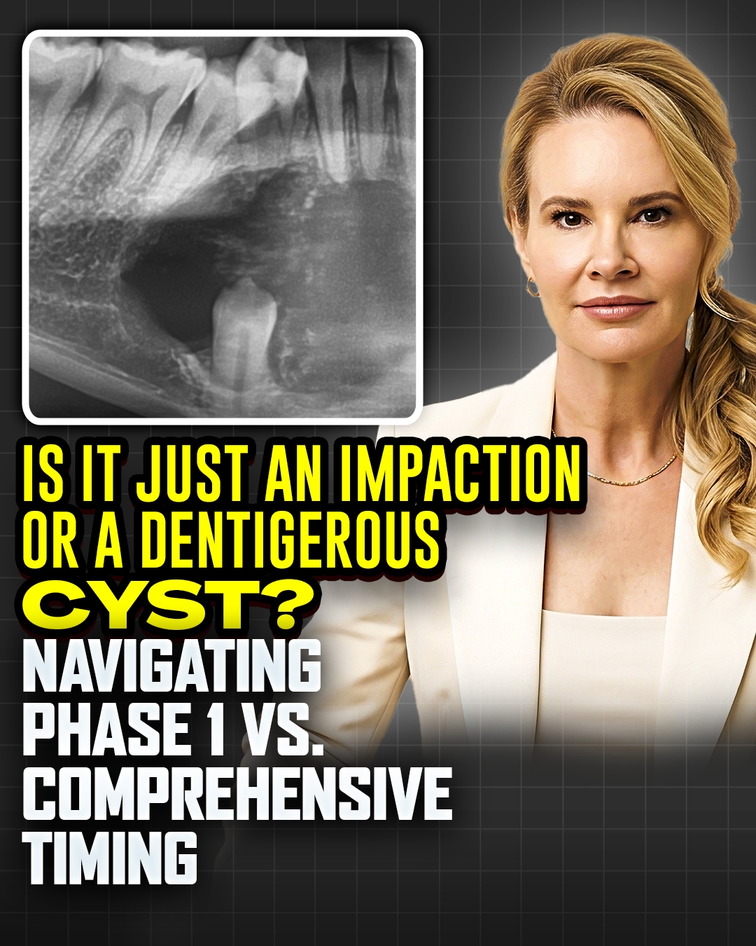
Canine Impactions and Dentigerous Cysts: Is Phase 1 Expansion Safe?
When managing impacted canines in Phase 1 treatment, our primary objective is typically straightforward: create space, perhaps use a 2×4 appliance, and utilize expansion to encourage auto-correction. However, discovering a dentigerous cyst—which looks like a fluid-filled balloon or bubble around the impacted crown—completely changes the clinical playbook.
While it is tempting to proceed with maxillary expansion to make room, the literature reveals three major reasons why applying active orthopedic force to a cystic maxilla is incredibly dangerous:
1. Pathological Cystic Expansion: Dentigerous cysts expand due to internal osmotic pressure. Orthopedic forces change the vascularity, suture stress, and fluid dynamics of the maxilla, which can cause the cyst to grow more aggressively, accelerating bone destruction around neighboring roots.
2. Root Displacement Risks: Tipping or translating incisors near a fluid-filled bone defect risks pushing healthy, moving roots directly into the cystic cavity. Teeth cannot move predictably or safely into a fluid defect; the bone must fill in first.
3. Acute Infection Risks: Maxillary expanders exert high forces on palatal shelves. If these forces cause microfractures in the thin cortical bone surrounding a large cyst, oral bacteria can enter, turning a benign sterile cyst into an acute maxillofacial infection.
The solution is not a highly invasive enucleation or premature surgical exposure if the canine is too high. Instead, the standard clinical protocol is decompression or marsupialization. An oral surgeon creates a small mucosal window to drain the fluid, dropping the pressure to zero so the body can naturally fill the defect with bone.
With the upcoming 2028 CODA airway mandates, more general practitioners are incorporating Phase 1 expansion into their practices. Understanding these pathological boundaries is vital to keeping treatment safe.